


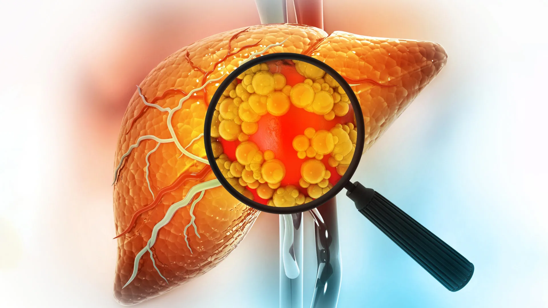
Low testosterone can contribute to several health problems, and an unhealthy diet may make some of those risks even worse. One major concern is metabolic dysfunction-associated steatotic liver disease (MASLD), a condition that affects approximately 40% of adult men worldwide and has become a significant global health issue.
Fatty liver, the earliest stage of MASLD, occurs when excess fat builds up in the liver. Several factors are associated with the condition, including obesity, type 2 diabetes, low testosterone levels, and excessive fructose consumption from sugary drinks and processed foods. However, scientists have not fully understood how these factors interact or whether their combined effects are more harmful than each one individually.
How Low Testosterone and Fructose Affect the Liver
To investigate this connection, a research team from the Graduate School of Agriculture, led by graduate student Hiroki Takahashi and Associate Professor Naoki Harada, examined how reduced testosterone and high fructose intake influence liver health.
The researchers used eight-week-old male mice and divided them into six experimental groups. Some underwent castration to lower testosterone levels, while others received a sham operation. The groups included Sham/Control, Sham/Fructose, Sham/Fructose + Antibiotics, Castration/Control, Castration/Fructose, and Castration/Fructose + Antibiotics.
To determine how these conditions affected the animals, the team analyzed liver cells and tissues, blood plasma, organic acids from the cecum, and samples of gut bacteria.
A Dangerous Combination for Fatty Liver
The results revealed that mice with reduced testosterone levels developed heavier livers when they consumed fructose. However, this increase in liver weight was smaller in mice that also received antibiotics.
When examined separately, low testosterone and fructose intake produced only modest changes in liver triglyceride levels. When combined, however, the two factors had a much stronger effect, significantly increasing fat accumulation and worsening fatty liver.
The researchers also identified changes in the composition of gut bacteria, altered liver gene activity, and elevated levels of pyruvate in the cecum among mice exposed to both reduced testosterone and fructose.
These findings suggested that the gut microbiome could play an important role in explaining why the combination was particularly harmful.
Gut Bacteria Reveal a Hidden Mechanism
Further experiments helped the researchers identify a possible biological explanation for the increased liver fat.
“Upon examining this mechanism, we found that changes in the gut microbiota led to increased levels of pyruvate within the intestine. Furthermore, experiments using mouse-derived primary hepatocytes revealed that pyruvate acts synergistically with fructose to promote neutral lipid accumulation in hepatocytes,” explained Takahashi.
In other words, changes in gut bacteria appeared to increase intestinal pyruvate, a compound involved in energy metabolism. Laboratory experiments with liver cells isolated from mice showed that pyruvate and fructose worked together to increase fat storage in those cells.
The discovery suggests that interactions among testosterone levels, dietary fructose, and gut bacteria may help explain how fatty liver develops and worsens.
Potential New Ways to Prevent Fatty Liver Disease
Understanding this relationship could eventually help researchers identify new approaches to preventing or treating fatty liver disease. In particular, targeting the processes through which pyruvate contributes to fat accumulation may offer promising possibilities.
“Going forward, we hope to clarify the mechanism by which pyruvate promotes triglyceride accumulation to pioneer the development of therapeutic drugs and establishment of preventive methods through dietary interventions,” added Professor Harada.
Although the findings come from experiments in mice and isolated mouse liver cells, they provide new insight into how hormonal changes and diet may interact to influence liver health.
The study was published in American Journal of Physiology-Endocrinology and Metabolism.
